OTOPLAN Software
Streamlined, Individualized Otological Surgery
With robust and accurate performance, OTOPLAN* has been developed in close collaboration between CASCINATION AG and MED-EL to unlock the full potential of individualized implantation. OTOPLAN’s unparalleled patient-specific 3D visualizations and measurement data at your fingertips will give you the power to efficiently perform individualized otological procedures with precision.
OTOPLAN also features automatic pre-op cochlear parameter measurements and electrode visualization, post-op implant detection, and allows you to leverage the future of fitting: anatomy-based fitting.
- Individualize otologic surgery & cochlear implant fitting maps
- Identify post-op electrode contact locations with an X-ray or CT scan
- Confirm electrode placement in scala tympani via CT image fusion
Automatic Inner Ear Reconstruction
Quicky reconstruct the full inner ear in 3D including the cochlea, scala tympani, scala vestibuli, round window membrane, bony overhang, semi-circular canals, and internal auditory canal.
Cochlear Implantation Planning
Perform virtual 3D scala tympani insertion to visualize insertion depth and frequency coverage of each MED-EL electrode. Plan the optimal placement of the implant housing and audio processor.
BONEBRIDGE Implantation Planning
Plan BONEBRIDGE placements that optimize implantations based on the individual anatomy of your patients with the newest version of OTOPLAN.
Post-Op Analysis
Visualize and perform a quality check of the insertion and lead management status. Determine postoperative location of each electrode contact with a plain X-ray or CT scan to export data for anatomy-based fitting.[ft]

Intuitive and Easy
With a guided workflow and an enhanced user interface, you can quickly master all of OTOPLAN’s features. OTOPLAN makes it easy to generate detailed 3D reconstructions and measure anatomical parameters in just a few quick steps.
Simple Data Management
Preview images, import, and export DICOM files quicker and more easily. For added convenience and compatibility, NRRD and NIfTI images can also be imported. The Moonliner tool allows for the automatic analysis of a batch of multiple patients with a few clicks.
Useful DICOM Viewing Tools
Perform measurements on DICOM images using 2D and 3D rulers, angular and freeform measurements, and a range of tools. Import, edit, play, and export fluoroscopy videos for complete patient records and presentations.
Optimized Guided Workflows
Step-by-step guidance highlights recommended actions and leads you through the clinical workflow.
Case Review Tools
OTOPLAN allows you to record your screen and voice, making it ideal for reviewing cases and teaching.

Electrode Visualization
Unique Combined View of Anatomical and Audiological Information
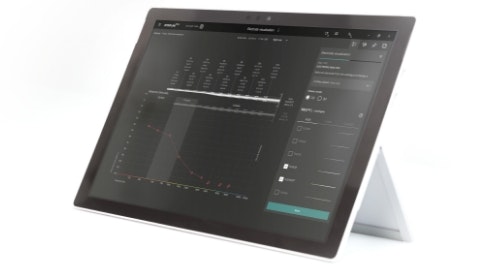
OTOPLAN gives you the power to take cochlear implant surgery planning to a whole new level. With the enhanced electrode visualization tool, you can combine anatomical and audiological information in one intuitive view. You can easily add preoperative data to a patient record and select desired cross-over frequencies for patients with residual hearing.
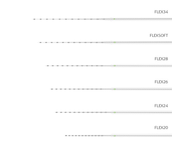
Even better, with OTOPLAN you can easily visualize how each electrode would fit each individual cochlea. Compare different electrode arrays and see detailed data for each individual electrode contact, including predicted angular insertion depth and tonotopic center frequency. Virtual 3D scala tympani insertion provides you with the scala ratio and other metrics, such as diameter, width, cross section, and volume.

3D EAR
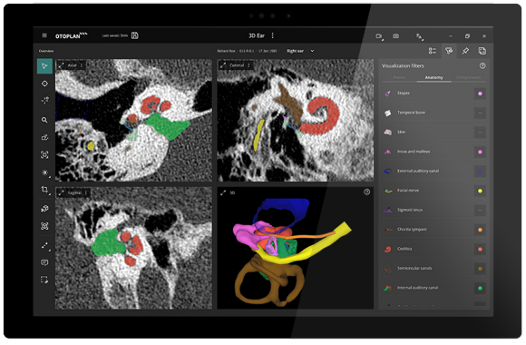
OTOPLAN gives you incredibly detailed 3D reconstructions of each patient’s anatomy in just seconds. Key anatomical structures—including the facial nerve, chorda tympani, sigmoid sinus, and the complete inner ear—are highlighted both in 3D and in each imaging plane.
Think of the possibilities: You can easily navigate through the temporal bone, chorda tympani, and facial nerve. Rotate 360° in any direction, zoom, or change transparencies. OTOPLAN makes it simple to see incredibly detailed 3D reconstructions.
- Automatic 3D inner ear
- Virtual trajectory planning
- Image fusion
Unparalleled Visualization
Instantly and seamlessly rotate and navigate through coronal, sagittal, and transverse anatomical planes to get an optimal 2D and 3D view of any anatomical structure in seconds.
3D Cochlea Reconstruction
Explore each individual cochlea via a detailed 3D reconstruction. This powerful tool can be especially helpful for assessing cochlear malformations. You can also export the model as an stl. file for 3D printing.
Bone and Skin Thickness Mapping
Perform automatic 3D reconstruction and thickness measurements of the skin and bone. Set a custom threshold and create a heatmap of skin or bone to see the distance to the dura or to air cells.
Virtual Trajectory Planning
You simply set the target point and then pivot the trajectory in any direction. This can be helpful for planning individualized surgical access to the cochlea for each patient.

Post-Op Analysis
OTOPLAN enhances the entire clinical workflow, including postoperative analysis and reporting. With postoperative imaging, you can easily confirm electrode insertion status and automatically generate detailed patient reports. OTOPLAN lets you automatically generate a report of a patient’s data, including a postoperative analysis.
With post-op CT scan data, OTOPLAN automatically determines the cochlear coordinate system and detects the electrode contacts, the electrode lead, and the implant housing. In addition, it provides visual feedback on the insertion status and lead management.
Using both CT and plain X-ray imaging, OTOPLAN allows you to quickly identify the postoperative angular insertion depth and tonotopic location of each individual electrode contact with just a few clicks. This real location data can be easily exported to our MAESTRO software for anatomy-based fitting.

Anatomy-Based Fitting
Our philosophy has always been to provide a closer match to natural hearing. Our unique full-length electrode arrays help enable more accurate place-pitch stimulation of the natural tonotopic map of the cochlea. Our unique FineHearing sound coding is designed to mimic natural rate coding in the second turn of the cochlea.
Now, with OTOPLAN and our MAESTRO fitting software, we are excited to reach closer to natural hearing than ever before with cochlear implants. Our default FineHearing frequency allocation is designed to follow the natural tonotopic map of the cochlea. However, we know that one size does not fit all. This is why we are always striving to provide a closer match for each individual patient.
With our new anatomy-based fitting tools, you can easily fine-tune the frequency map to be closer to the natural map of each individual cochlea. By combining plain X-ray or CT imaging data from OTOPLAN with MAESTRO, you can now quickly assign center frequencies based on the actual anatomical location of each electrode contact.
Discover More

Does Your Clinic Have the Newest Version of OTOPLAN?
Make sure you're getting the most out of OTOPLAN with the newest version. It allows you to identify post-op electrode contact locations with an X-ray or CT scan and confirm post-op electrode placement in the scala tympani via CT image fusion, along with all of the powerful tools listed below.
| Feature | Newest Version (2024 release) |
| Data management | Supports DICOM, NRRD, and NIfTI files |
| 2D and 3D measurement tools | Linear ruler, polygonal ruler, spline ruler, freeform measurement, angular measurement, and closest distance between 3D structures |
| Automatic 3D inner ear reconstruction | Cochlea, scala tympani, scala vestibuli, round window, bony overhang, semi-circular canals, and internal auditory canal |
| Cochlear parameter measurements | Automatic |
| Virtual electrode insertion | 2D and 3D |
| Implant placement planning | Cochlear implants and BONEBRIDGE |
| 3D temporal bone reconstruction and thickness mapping | Automatic |
| Skin reconstruction and thickness mapping | Automatic |
| Bilateral implant housing and audio processor placement planning | Cochlear implants |
| 3D Ear | Includes the facial nerve, chorda tympani, sigmoid sinus, ossicles, and the complete inner ear with scala tympani |
| Automatic detection in post-op analysis with CT scan | Cochlear coordinate system, electrode contacts, electrode lead, and implant housing |
| Manual detection in post-op analysis with plain X-ray | Electrode contacts |
| Image fusion | CT & MRI |
| Moonliner image batch analysis tool | Automatic, preoperative and postoperative |
| Fluoroscopy video editor | Import, edit, play, and export |

Contact Us
Ready to further individualize and streamline otological surgery at your clinic with OTOPLAN?
Would you like to upgrade to OTOPLAN's newest version?
Fill in the contact form below. We’ll connect you to our local MED-EL clinical support experts.

SYNCHRONY 2
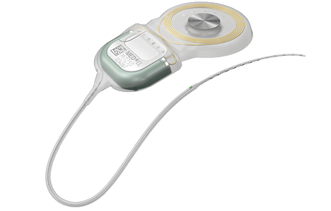
See why individualized electrode arrays, safe access to MRI, and a streamlined central electrode lead for exceptional surgical handling make SYNCHRONY 2 your new favorite cochlear implant.
Discover More
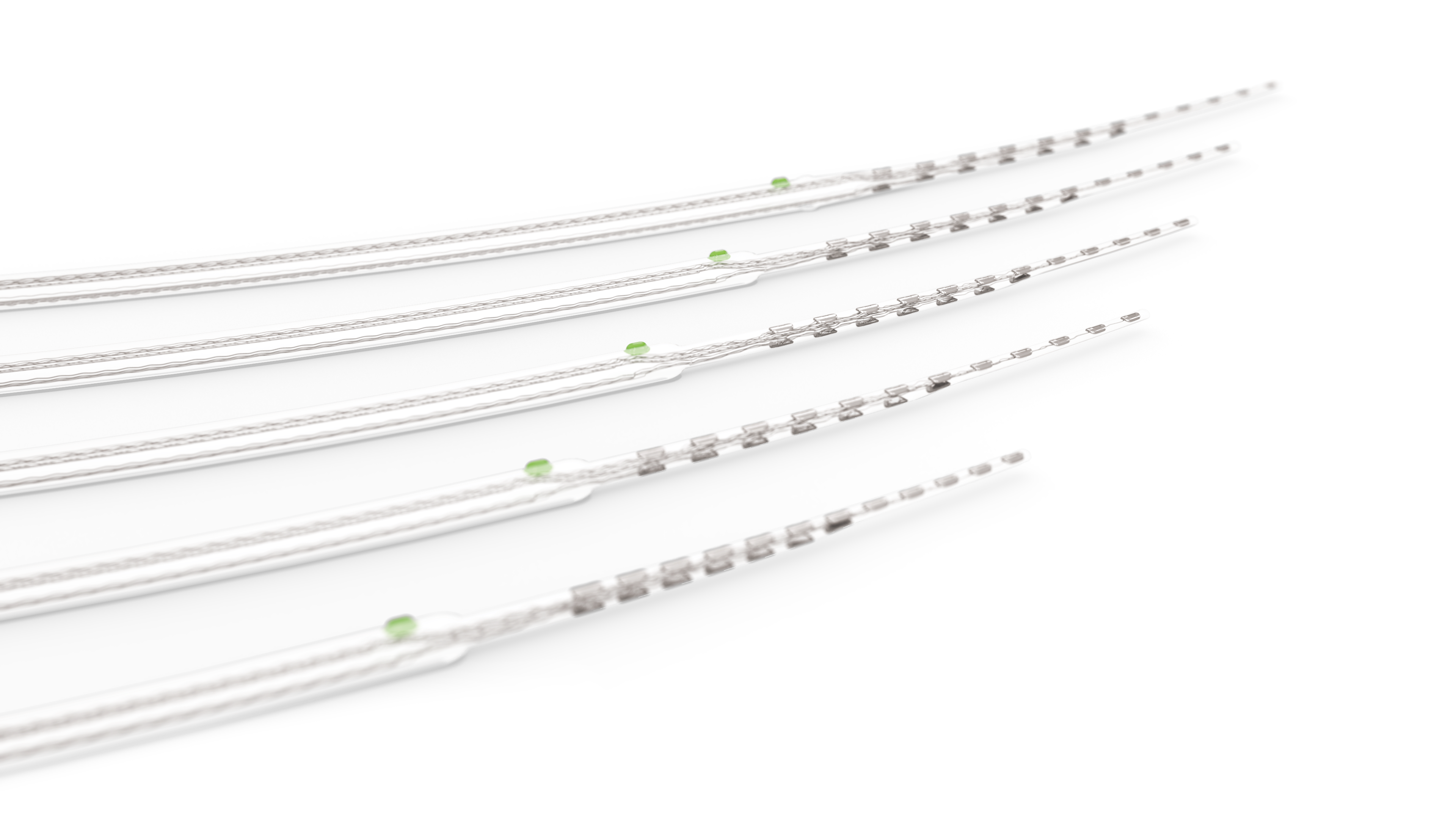
Electrodes
Find out what sets MED-EL electrode arrays apart from any other design to enable a closer to natural sound quality that no other cochlear implant can match.
*OTOPLAN is a product of CASCINATION AG.
