MED-EL Cochlear Implant Electrode Arrays

electrode

Above all else, the purpose of a cochlear implant is to create the best possible communication between the electrode array and the delicate neural structures of cochlea. All of the incredible technology in our cochlear implants flows across this intricate bridge between technology and nature.

How do we provide the most natural hearing possible with a cochlear implant? By engineering our electrode arrays to most closely match the precise natural design and function of the cochlea.

Our incredibly flexible electrode arrays help preserve the delicate structures in the cochlea, enabling atraumatic scala tympani insertion all the way to the apical region. This allows our full-length electrode arrays to provide two full turns of Complete Cochlear Coverage, which maximizes the natural tonotopic stimulation range.[ft][ft][ft][ft][ft][ft][ft]

Finally, our unique variable-rate FineHearing sound coding is the only sound coding that uses two types of sound coding to mimic both tonotopic stimulation and temporal phase-locking in natural hearing for a sound quality that no other cochlear implant can match.[ft][ft][ft][ft][ft][ft]

All together, these unique MED-EL technologies enable our electrodes to provide the closest to natural stimulation all along the cochlea for the best hearing outcomes.

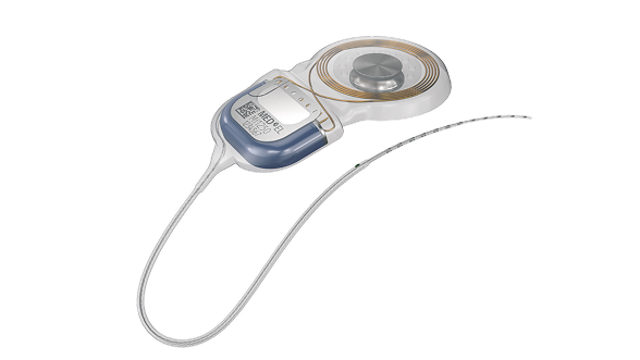
But one size does not fit all—which is why we have individualized electrode arrays to best fit each unique cochlea. By enabling the closest possible match between electrode array and each individual cochlea, we’re taking our cochlear implants closer to natural hearing than ever before.[ft][ft]

  • Preserving Cochlear Structures

Our incredibly flexible electrode arrays help preserve the delicate structures in the cochlea, enabling atraumatic scala tympani insertion all the way to the apical region.[ft][ft][ft][ft][ft][ft][ft]

  • Covering the Whole Cochlea

Only MED-EL offers electrode arrays long enough to cover up to two full turns of the cochlea for better hearing performance and more natural sound quality.[ft][ft][ft][ft][ft][ft]

  • Matching Each Cochlea

With our comprehensive electrode portfolio, you can easily choose the ideal array to match each individual cochlea.[ft][ft]

  • Both Types of Natural Sound Coding

Only MED-EL provides a combination of both types of sound coding for the closest to natural sound perception.[ft][ft][ft][ft][ft][ft]

MED-EL Cochlear Implant

Structure Preservation

First, do no harm. Even in profound sensorineural hearing loss, the cochlea is full of living neural structures—a deaf ear is not a dead ear. These intricate natural structures are a fraction of the diameter of a human hair, making ultra-flexible electrode arrays necessary.

For more than 25 years, we’ve worked to create the most atraumatic cochlear implant electrode arrays possible. Our free-fitting arrays are uniquely engineered to deliver unparalleled flexibility, making them the most atraumatic cochlear implant electrode arrays available.[ft]

Our incredibly flexible lateral wall arrays gently adapt to the shape of each individual cochlea to protect the delicate natural structures. Multiple recent studies have shown only MED-EL arrays enable up to 100% scala tympani insertions, which enables significantly better hearing outcomes. Our gentle arrays are also ideal for supporting hearing preservation, for combined electric-acoustic stimulation.

Free-Fitting Arrays

Our free-fitting arrays gently flex to adapt to the unique shape of each cochlea while bringing electrode contacts close to neural targets.

Wave-Shaped Wires

Uniquely shaped flexible wires bend more easily, which helps reduce rigidity and insertion forces in comparison to straight-wire designs.[ft]

Optimal Contact Spacing

Our design offers an ideal combination of mechanical flexibility and channel spacing to reduce insertion force and avoid overlapping stimulation channels.[ft][ft]

Round Window Insertion

With no need for an insertion stylet, our arrays can be atraumatically inserted through the round window into the scala tympani, avoiding drilling trauma from a cochleostomy.[ft]

FLEX-Tip Technology

FLEX arrays feature a tapered, rounded tip that gently glides along the scala tympani during insertion, avoiding damage to the basilar membrane and delicate structures.[ft]

Scalar Deviation

The foundation of structure preservation is electrode placement fully in the scala tympani. If an electrode array deviates into the scala vestibuli, then the perilymph and endolymph would likely mix, destroying the endocochlear potential and impairing essential nerve function.[ft][ft][ft][ft]

However, the scala tympani is separated from the scala vestibuli by incredibly delicate structures that measure only a few microns in diameter. Stiff precurved arrays are far more likely to penetrate the basilar membrane and cross into the scala vestibuli. This issue is also possible with stiff straight arrays.

Only MED-EL’s ultra-flexible free-fitting arrays are proven to enable reliable scala tympani placement. By gently flexing away from delicate structures, our FLEX arrays enable atraumatic deep insertion into the cochlea without harming the basilar membrane or other essential neural structures.[ft][ft][ft][ft][ft][ft][ft][ft]

"It is important that electrodes do not perforate the structures of the cochlea. If the electrode deviates into the scala vestibule, it usually damages the scala media . . . the fluid in the cochlea mixes with the fluid surrounding the ganglion cells, meaning that a toxic inflammation in the cochlea will spread to the nerve." – Prof. Helge Rask-Andersen

Literature Search Report (2020), Data on File, MED-EL n=1,399
O´Connell et al. 2016

Round Window Insertion

The round window provides direct access to the scala tympani, making it the ideal route for atraumatic insertion. This avoids the potential trauma of drilling a cochleostomy, as well as the risk of scala vestibuli placement.[ft][ft]

With a 0.8–1.3 mm basal diameter, our arrays are designed for atraumatic round window insertion. And with no need for an insertion stiffener or stiff sheath to be inserted in the cochlea, there is minimal disturbance to the delicate structures at the base of the cochlea.

Electrode FLEX

Atraumatic Deep Insertion

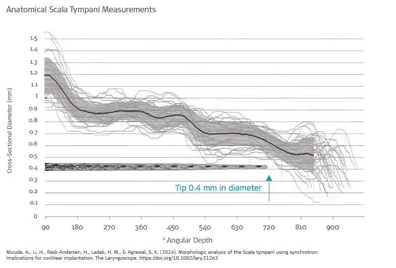
As the cochlea nears the apex, the cross section of the scala tympani tapers in diameter to approximately 0.7 mm at 720°. This is why it is important to ensure the array can safely fit in the cochlear duct. However, with other thin arrays, the electrode array only covers the basal turn, where a reduced diameter is not necessary.

Our free-fitting FLEX arrays use a tapered FLEXTip for the apical 5 electrode contacts, making FLEX series the thinnest full-length electrode arrays available. With a tip diameter of 0.4 x 0.5 mm or less, the FLEXTip can be safely and reliable placed in the second turn of the scala tympani without harming the basilar membrane.


Electrode in Cochlear

Tip Fold-Over

With pre-curved electrode arrays, the tip of the electrode array can also become lodged and fold over upon itself. This tip fold-over complication can seriously compromise the interface between the electrode contacts and the cochlea, and often limits coverage to less than ¾ of a single turn in the cochlea.[ft][ft][ft][ft]

With MED-EL arrays, tip fold-over is rare, with MED-EL having the lowest reported rate of tip fold-over.[ft][ft][ft][ft][ft][ft][ft][ft][ft][ft][ft][ft] The rounded FLEXTip of our arrays gently adapts to the anatomy of each individual cochlea, so there is a very minimized risk of the tip becoming lodged or folding over. In an extensive review of more than 1,500 implantations, tip-fold over repeatedly occurred with pre-curved and rigid straight arrays, but was not encountered with any of the 700+ MED-EL arrays.[ft]

Tip Fold-Over
Gabrielpillai et al. 2018

Hearing Preservation

Preserving the natural function of the cochlea is especially important for patients with residual hearing. If an array deviates into the scala vestibuli, all residual hearing would likely be permanently lost. Stiff straight and pre-curved arrays are both far more likely to cause loss of residual hearing.[ft][ft][ft][ft][ft][ft][ft]

Our ultra-flexible arrays enable a superior hearing preservation rate for combined electric-acoustic stimulation. Long-term post-operative residual hearing is a useful indication that the natural anatomy and physiology of the cochlea has been preserved. This also offers a more intact foundation of neural structures for future therapies.[ft] [ft][ft][ft][ft]

Closest to Natural Hearing

Closest to Natural Hearing

In the cochlea, natural frequency response is intricately ordered in on a logarithmic scale across two full turns. This tonotopic place-pitch frequency response along the whole cochlea allows frequency mapping along a clear, logical path.[ft]

How do we provide the closest to natural hearing with our electrode arrays? By aligning our electrodes as closely as possible to the natural tonotopic place across the whole cochlea. What does more natural sound quality mean for you patients? More natural music enjoyment, much better hearing in everyday life, and the best possible hearing experience with their CI.[ft][ft][ft][ft][ft]

Complete Cochlear Coverage

In natural hearing, the nerve fibers of the spiral ganglion are tonotopically organized and extend two full turns in the cochlea. If an electrode does not cover up to two turns (540-720°) of the cochlea, it cannot stimulate these nerve fibers and cannot provide accurate pitch-place match.[ft]

Other arrays only cover the basal turn of the cochlea (~360-400°), limiting the stimulation range and leaving half of the cochlea unused. Only MED-EL offers long, flexible electrode arrays up to 34 mm in length to provide up to two full turns (720°) of cochlear coverage, reaching the apical region.[ft][ft]


Natural Tonotopic Match

If an electrode only covers half the cochlea, frequencies below ~1,000 Hz can’t be stimulated at the right place, so these frequencies would be shifted upwards. This causes an unnatural sound quality that recipients describe as “robotic”, “tinny”, “echoey”, or “mechanical”. Adapting to this pitch-place mismatch can take years for even partial improvement.[ft][ft][ft]

With our long arrays that offer up to 720° of angular insertion depth, the electrode array can align with the natural tonotopic map of the basilar membrane and spiral ganglion all along the cochlea. This means a 150 Hz tone can be stimulated through the same nerve fibers as a natural 150 Hz tone, instead of being artificially transposed to the tonotopic cochlear place of a 1,000 Hz tone. This more accurate pitch match enables a much more natural hearing experience for your patients. [ft][ft][ft][ft][ft]

Data on file, calculated from mean cochlear parameters

Hearing Performance

Why stop at closer to natural sound quality? By using more of the natural cochlear potential, our long arrays enable significantly better hearing performance. This benefit is especially useful in challenging listening environments, like following a conversation in a noisy restaurant. Having a closer match from day one can also support faster improvements in hearing ability.[ft][ft][ft]

Buechner et al. 2017

FineHearing: Natural Sound Coding

In nature, the cochlea uses two types of sound coding. Place-pitch tonotopic coding is used along the whole frequency range. In addition to place-pitch coding, low-frequency sounds are also coded by rate. With this time-coding, the frequency of nerve signals is synchronized to the frequency of the sound below ~1000 Hz.

That’s why we created the only cochlear implant sound coding that mimics the natural time-coding for low frequencies and provides place-pitch match throughout the cochlea. By mimicking natural sound coding on each electrode contact, FineHearing provides much more natural sound quality.[ft][ft][ft][ft]

“[MED-EL’s] place dependent stimulation rates allow thus far unparalleled restoration of tonotopic pitch perception.” – Rader et al. 2016


Individualized Cochlear Implants

Individualized
Cochlear Implants

When it comes to electrode arrays, one size doesn’t fit all. In fact, there’s a wider range of variation in cochlear duct lengths than shoe sizes—up to 50% difference between two cochleae. That’s why we designed our FLEX Series arrays to provide the optimal length for the full range of cochlear anatomies.[ft][ft]

With six FLEX arrays available in sizes between 20–34 mm, you can achieve complete cochlear coverage and full electrode insertion for each patient. And with OTOPLAN, the intuitive surgical planning software, you can easily optimize the electrode array based on the measurements of each individual cochlea.

Our philosophy is straightforward: Adapt the implant to your patient, instead of adapting your patient to the implant.

“This is a huge advantage that we can individualize the choice of these electrode lengths. We know there’s a huge variation in sizes from cochlear lengths. The organ of Corti, the cochlear duct length varies from 25– 36 millimeters, really this is a 50% change, from one patient to another patient.” – Prof. Paul van de Heyning


Electrodes

OTOPLAN: Powerful Visualization

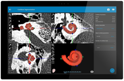
OTOPLAN is fully portable and tablet-based, giving you exceptional mobility and flexibility. The responsive touch-screen controls put the power in your fingertips. OTOPLAN gives you incredibly detailed 3D reconstruction of the anatomical structures in just seconds, including the stapes and the cochlear basal turn. Key anatomical structures—including facial nerve, chorda tympani, and ossicular chain, and basal turn of the cochlea—are highlighted both in 3D and in each imaging plane.*


OTOPLAN

Optimal Cochlear View

With OTOPLAN, you can easily rotate the imaging planes to achieve an ideal oblique view of the cochlea. An optimal cochlear view enables more accurate and consistent measurements, for better surgical quality and electrode selection.

With just a few clicks in the guided workflow, you can accurately measure cochlear diameter and cochlear height. OTOPLAN instantly calculates cochlear duct length using a precise algorithm.


Surface Pro

Electrode Visualization Audiogram

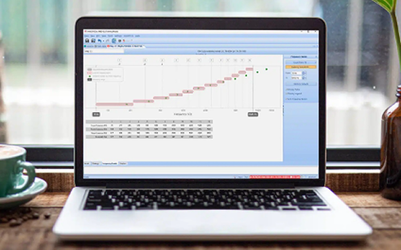
Even better, OTOPLAN’s electrode visualization tool lets you combine the anatomical and audiological information in one intuitive view. Now, you can easily visualize how each MED-EL electrode array fits each cochlea.

In fact, you can see angular insertion depth, tonotopic frequency and % cochlear coverage for each electrode contact on any array, as well as integrated audiogram data for each patient. OTOPLAN makes it easier than ever before for you to choose the ideal electrode array for each individual patient.


Electrode Visualization FLEX 34
MED-EL

Why MED-EL:
A Trusted Partner

For more than 30 years, MED-EL has been a trusted partner and innovation leader in hearing implants. We’re here to work together with you, and we’re committed to providing outstanding service and support for our professional partners.

With the most advanced cochlear implant systems, we offer the best hearing experience for your patients and the best clinical experience for you.


  • Closest to Natural Hearing
  • Superior Ease-of-Use
  • True Peace of Mind

Contact

Ready to learn more about MED-EL cochlear implants?
Fill out our simple contact form and we’ll get in touch with you.

Personal Information

Clinic Information

Comment*

We will treat your information with respect. For more information about our privacy practices please visit our privacy policy page. By clicking below, you agree that we may process your information in accordance with these terms.

Your request and contact details will be forwarded to the closest MED-EL office or certified partner in your country. MED-EL will use the information you provide on this form to be in touch with you and to provide updates and marketing.

More From MED-EL

How would you like to hear from us?

< Previous Step
easyMRI 
007_MSY2_v037_SY2P_SVec_bk-bg_per2

SYNCHRONY 2

See why individualized electrode arrays, safe access to MRI, and a streamlined central electrode lead for exceptional surgical handling make SYNCHRONY 2 your new favorite cochlear implant.


Discover More 

OTOPLAN

With intuitive user interface and powerful 3D visualization, OTOPLAN is the ideal surgical planning software for otological procedures.

Discover More


Otoplan

* OTOPLAN software is a product of CAScination AG.

Electrode Arrays


FLEX Series

The softest and most flexible electrode arrays, designed for Structure Preservation and Complete Cochlear Coverage. Featuring 19 active and physical platinum electrode contacts and FLEX-tip technology for atraumatic insertion.


FLEX34

  • 28.6 mm stimulation range
  • Diameter at basal end: 1.3 mm
  • Dimensions at apical end: 0.5 x 0.4 mm

FLEXSOFT

  • 26.4 mm stimulation range
  • Diameter at basal end: 1.3 mm
  • Dimensions at apical end: 0.5 x 0.4 mm

FLEX28

  • 23.1 mm stimulation range
  • Diameter at basal end: 0.8 mm
  • Dimensions at apical end: 0.5 x 0.4 mm

FLEX26

  • 20.9 mm stimulation range
  • Diameter at basal end: 0.8 mm
  • Dimensions at apical end: 0.5 x 0.3 mm

FLEX24

  • 20.9 mm stimulation range
  • Diameter at basal end: 0.8 mm
  • Dimensions at apical end: 0.5 x 0.3 mm

FLEX20

  • 15.4 mm stimulation range
  • Diameter at basal end: 0.8 mm
  • Dimensions at apical end: 0.5 x 0.3 mm

FORM Series

Designed specifically for malformed cochleae and for instances where leakage of cerebrospinal fluid (CSF) is expected. Featuring 24 active and physical platinum electrode contacts and SEAL technology designed to aid closing of the cochlear opening.


FORM24

  • 18.7 mm stimulation range
  • Diameter at basal end: 0.8 mm
  • Diameter at apical end: 0.5 mm

FORM19

  • 14.3 mm stimulation range
  • Diameter at basal end: 0.8 mm
  • Diameter at apical end: 0.5 mm

CLASSIC Series

Features 24 active and physical platinum electrode contacts.

STANDARD

  • 26.4 mm stimulation range
  • Diameter at basal end: 1.3 mm
  • Diameter at apical end: 0.5 mm

MEDIUM

  • 20.9 mm stimulation range
  • Diameter at basal end: 0.8 mm
  • Diameter at apical end: 0.5 mm

COMPRESSED

  • 12.1 mm stimulation range
  • Diameter at basal end: 0.7 mm
  • Diameter at apical end: 0.5 mm